How Myasthenia Gravis Weakens the Neuromuscular Junction
See how acetylcholine, its receptors, and autoantibodies alter signaling from nerve to muscle. Symptoms alone cannot establish a diagnosis, and worsening breathing or swallowing needs urgent assessment.
Publication & corrections: infostoa · Editorial standards
Individual author and specialist reviewer information is not provided for this article.
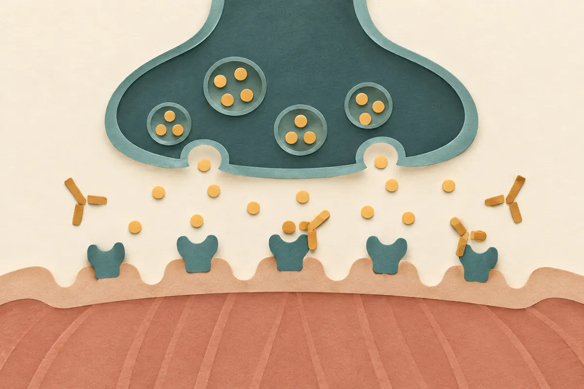
Myasthenia gravis is an autoimmune disorder in which signaling from nerve to muscle becomes less reliable at the neuromuscular junction. It can cause weakness or easy fatigability in the eyes, face, limbs, swallowing muscles, or breathing muscles. The mechanism is not simply that the nerve stops releasing acetylcholine: immune attack targets receptors and related structures on the muscle side of the junction.
Normal signaling crosses a tiny gap
When an electrical impulse reaches a motor nerve ending, the nerve releases acetylcholine into the synaptic cleft. The transmitter binds nicotinic acetylcholine receptors on the muscle membrane. If enough receptors respond, the muscle membrane reaches the electrical threshold that initiates contraction. Release, diffusion, receptor binding, and transmitter breakdown all contribute to this sequence.
The junction normally has a safety margin: a nerve impulse produces a response large enough to activate the muscle. Myasthenia reduces that margin. A smaller effective response can still sometimes trigger contraction, but repeated activity may expose the limited reserve. This helps explain fluctuating weakness.
Autoantibodies reduce the junction’s reserve
Many people with myasthenia gravis have antibodies against the acetylcholine receptor, or AChR. Depending on the antibody and immune process, receptors may be blocked, removed from the membrane, or the postsynaptic folds may be damaged through complement activity. Fewer functioning receptors make it harder for the same acetylcholine signal to produce a muscle response.
Other patients have antibodies against proteins such as muscle-specific kinase (MuSK), which helps organize receptors at the junction, or LRP4. These are not interchangeable mechanisms in every detail, and antibody patterns vary. A negative test for one antibody does not by itself settle whether myasthenia is present; clinicians interpret tests alongside symptoms and other studies.
The thymus is associated with the immune system and may be abnormal in some patients, including enlargement or a thymoma. Its precise role is not fully understood, and a thymus finding does not explain every case. The mechanistic account should therefore avoid a single universal cause.
Repeated activity can reveal fatigable weakness
Common patterns include drooping eyelids, double vision, changes in facial expression, difficulty chewing or speaking, trouble swallowing, and weakness in the arms, legs, or neck. Symptoms can worsen with repeated use and improve after rest, although the pattern differs across individuals. Some people have weakness limited mainly to eye muscles; others have generalized involvement.
At a junction with less reserve, successive nerve impulses may be less likely to produce a full muscle response. This offers one explanation for fatigability. It does not mean that ordinary tiredness proves myasthenia: fatigue and weakness have many causes, and a symptom description is not a diagnosis.
A brief record of when weakness occurs, the activity involved, whether rest changes it, and which functions are affected—such as vision, speech, or swallowing—may help a clinician understand variation. A log is not a diagnosis, and rapidly worsening breathing or swallowing should not wait while someone gathers more entries.
Expert perspective: diagnosis combines the clinical picture and tests
A clinician considers the distribution and fluctuation of symptoms, examination findings, and tests. Blood testing may look for AChR or other relevant antibodies. Repetitive nerve stimulation and single-fiber electromyography can assess transmission at the junction; imaging may be used to examine the thymus. No single test answers every question in every patient, and negative antibody results do not automatically exclude the disorder.
Infection, surgery, and some medicines can worsen symptoms in some people. Do not stop a prescribed medicine or change a dose based on a general list; discuss a possible trigger with the prescribing clinician, because the risk depends on the drug and the person’s condition.
Antibody results inform the mechanism of impaired neuromuscular transmission, but do not directly measure a person’s strength or severity. A negative test is not conclusive on its own. Clinicians combine symptoms over time, examination, blood tests, and electrophysiology.
Treatment choices follow an individual assessment
Acetylcholinesterase inhibitors slow the breakdown of acetylcholine, leaving more transmitter available at the junction. Other treatment options may affect immune activity or rapidly change circulating antibodies; thymus surgery is relevant for selected patients. Which option is appropriate depends on the antibody pattern, symptom distribution, severity, age, other conditions, and specialist assessment. A mechanism overview cannot select a treatment or dose for an individual.
Sudden or worsening difficulty breathing or swallowing can be an emergency, including a possible myasthenic crisis. Seek urgent medical care rather than relying on an article or waiting to see whether rest is enough. For nonurgent questions, a neurologist or neuromuscular specialist can interpret symptoms and tests together.