Ischemic Stroke: The Penumbra and Reperfusion Assessment
In ischemic stroke, the infarct core and surrounding tissue still at risk are different tissue states. Reperfusion may rescue some viable tissue, but candidacy is determined through imaging and clinical assessment.
Publication & corrections: infostoa · Editorial standards
Individual author and specialist reviewer information is not provided for this article.
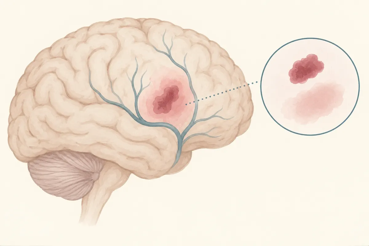
In ischemic stroke, not all tissue affected by reduced blood flow is injured to the same degree. The National Institute of Neurological Disorders and Stroke (NINDS) distinguishes an irreversibly damaged core from tissue that remains viable but at risk. Professional clinical material describes the surrounding region as the ischemic penumbra. Reperfusion may rescue tissue that is still viable, but the term alone cannot determine whether a person is a candidate for treatment.
What the core and penumbra describe
When a vessel is blocked or blood flow falls, the brain receives less oxygen and fewer nutrients. The most severely underperfused core may progress to irreversible injury, while some surrounding tissue may retain function for a time despite inadequate flow. Collateral circulation through nearby vessels is one factor that can support this tissue. The boundary is not a fixed line: it varies with the location and extent of the blockage, collateral flow, elapsed time, and the person's condition.
NINDS explains that viable but threatened tissue may surround the core after an ischemic stroke and that reperfusion is important for rescuing tissue that remains viable. The Merck Manual Professional Edition likewise describes penumbral tissue around more severely ischemic areas. This is a biological model, not a way to infer the size of a core or the presence of a penumbra from symptoms or a clock at home.
Why imaging and examination matter
A clinical team uses neurologic symptoms and examination, together with imaging such as CT or MRI, to evaluate bleeding, vessel blockage, injury, and blood flow. The Merck Manual describes perfusion CT or MRI as methods for assessing infarcted tissue and underperfused tissue at risk. Comparing diffusion and perfusion imaging may also be used in specific clinical contexts. No single imaging value guarantees an outcome or proves that a particular intervention will help.
The presence and extent of a penumbra require specialist interpretation. Vascular anatomy, collateral flow, scan timing, changing symptoms, and other conditions may affect the assessment. Having an image does not make the same drug or procedure appropriate for every patient. A stroke team weighs the evidence about potentially viable tissue alongside the likely benefits and risks of treatment.
Why a single time number cannot determine reperfusion treatment
Thrombolytic therapy and endovascular thrombectomy both aim to restore blood flow, but their selection criteria and risks differ. Professional sources describe potential benefit for selected patients while emphasizing clinical findings and imaging. Treatment decisions can depend on onset history, vessel location, brain imaging, whether deficits are disabling, bleeding risk, and the capabilities of the treating center.
Time is an important part of evaluation, but a threshold from one guideline, country, or publication period should not be reduced to a universal “golden window.” It neither guarantees eligibility nor serves as a self-screening rule. Current recommendations and local protocols may differ. This article is a mechanism overview; it does not provide a current treatment window, drug choice, dose, or clinical protocol.
Use the penumbra concept safely
The practical point is that continued loss of blood flow can injure tissue that is still at risk, so rapid emergency assessment matters. Sudden weakness or numbness of the face, arm, or leg, difficulty speaking, or an abrupt change in vision or balance can be signs of stroke. Contact local emergency services immediately rather than calculating a time window at home. Do not delay evaluation if symptoms improve.
A companion can note when symptoms began or when the person was last known well, along with medicines and relevant history, to help the emergency team. Those details assist clinical assessment; they do not reveal a penumbra without imaging. Treatment and prognosis must be discussed with clinicians. An online mechanism overview cannot replace examination or hospital imaging.
Expert perspective: the penumbra is not a self-triage chart
NINDS explains the core, at-risk tissue, and rationale for reperfusion; the Merck Manual Professional Edition discusses collateral flow and perfusion imaging. These sources support a mechanism and specialist-assessment context, not an individual determination of tissue viability or treatment. Use local emergency services for suspected stroke and ask the treating team about applicable criteria.