HFrEF: Neurohormonal Compensation and Ventricular Remodeling
Learn what reduced ejection fraction means and how sympathetic and RAAS responses can support circulation initially yet add long-term cardiac strain. Not personal medical advice.
Publication & corrections: infostoa · Editorial standards
Individual author and specialist reviewer information is not provided for this article.
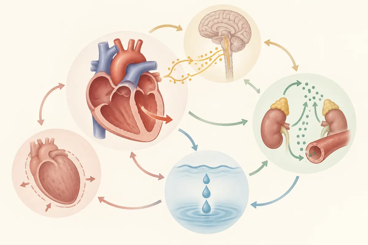
Heart failure with reduced ejection fraction (HFrEF) describes impaired left ventricular contraction with a lower share of filled blood ejected on each beat. The body activates neural and hormonal responses to preserve circulation, but sustained compensation can increase cardiac workload and fluid congestion. This article traces those mechanisms. An ejection-fraction number alone cannot establish an individual diagnosis or treatment plan.
What ejection fraction tells you
The Merck Manual Professional Edition describes HFrEF as left ventricular systolic dysfunction with an ejection fraction of 40% or less. Ejection fraction is the proportion of blood in the ventricle at filling that leaves with a beat. Heart failure—when the heart cannot supply enough blood for the body—can also involve inadequate filling, not only weak contraction. Ejection fraction therefore does not, by itself, describe every symptom, cause, or degree of congestion.
The National Heart, Lung, and Blood Institute (NHLBI) explains that heart failure occurs when the heart cannot pump enough blood for the body’s needs. This can happen when the heart is too weak to pump properly or cannot fill adequately. Fatigue, breathlessness, and fluid buildup can occur, but clinicians diagnose the condition using history, examination, imaging, and blood tests. The HFrEF classification threshold and interpretation for one person are separate questions.
Compensation initially supports circulation
When cardiac output falls, baroreflexes increase sympathetic activity and reduce parasympathetic influence. A faster heart rate, stronger contraction, and narrowed vessels can help maintain blood pressure and organ perfusion in the short term. If kidney blood flow falls, the renin-angiotensin-aldosterone system (RAAS) can also become active. Angiotensin II constricts vessels, while aldosterone promotes sodium and water retention, helping restore circulating volume.
These responses compensate; they do not remove the cause of injury. More returning blood increases ventricular filling load, and greater vascular resistance raises the load against which the heart ejects. If renal underperfusion persists, RAAS stimulation may continue. The pathways interact, but their degree and clinical effects are not identical in every patient.
How persistent signals can contribute to congestion and remodeling
Retained fluid can raise filling pressure and contribute to congestion in the lungs or peripheral tissues. Ongoing sympathetic stimulation increases heart rate and oxygen demand, while vasoconstriction increases resistance to ejection. These loads and hormonal signals can contribute to left ventricular dilation, changes in shape, fibrosis, and other adverse remodeling. A response that initially supports circulation can thus become part of a cycle that worsens cardiac function over time.
This is a general mechanism, not a forecast of one person’s course. The underlying cause, cardiac structure, kidney function, rhythm, other conditions, and timing of measurements all affect the clinical picture. A change in symptoms or new breathlessness or swelling should be assessed clinically rather than interpreted by comparing ejection-fraction values alone.
Ejection fraction is a measure of ventricular function, often assessed with cardiac imaging, and its meaning depends on the clinical context and timing. A measurement during congestion may not describe the same state as one obtained after the condition changes. Symptoms and functional limitations also differ among people with similar values. A borderline value does not by itself settle whether heart failure is present; clinicians interpret it with cardiac structure, symptoms, and other tests.
Separate a mechanism from an individual course
The same neurohormonal response can have different consequences depending on the cause of heart failure, which ventricles are involved, kidney function, and fluid status. A mechanism model shows how signals across organs connect; it does not quantify which pathway matters most for one person. If measurements or symptoms differ from expectations, discuss the clinical context with a clinician rather than drawing a conclusion from this overview alone.
Expert perspective: do not turn a pathway into a personal prescription
The professional overview helps explain why short-term compensation and long-term strain can coexist. A mechanism description is not an instruction to start, stop, or select a drug or dose. Clinicians weigh the cause, test results, kidney and electrolyte status, blood pressure, symptoms, and other medicines when choosing care. NHLBI likewise describes diagnosis as a combination of medical history, examination, imaging, and blood tests.