How Neuronal Hyperexcitability and Epileptiform EEG Discharges Arise
Learn how excitation, inhibition, and network synchrony relate to epileptiform EEG patterns, and how human-tissue findings differ from broader network models.
Publication & corrections: infostoa · Editorial standards
Individual author and specialist reviewer information is not provided for this article.
The short answer: epileptiform discharges reflect coordinated networks, not one “overactive” cell
Neuronal hyperexcitability does not mean that every neuron in the brain is constantly firing too much. It describes conditions in which the balance and timing of cellular inputs, intrinsic firing properties, and connected circuits can make particular networks more likely to produce abnormal synchronized activity. When electrical activity across many neurons becomes coordinated, an electroencephalogram (EEG) may record an interictal spike or a longer ictal discharge. The familiar shorthand of “too much excitation and too little inhibition” can be useful, but it is not a universal explanation for the many epilepsies, which arise from different causes and circuits.
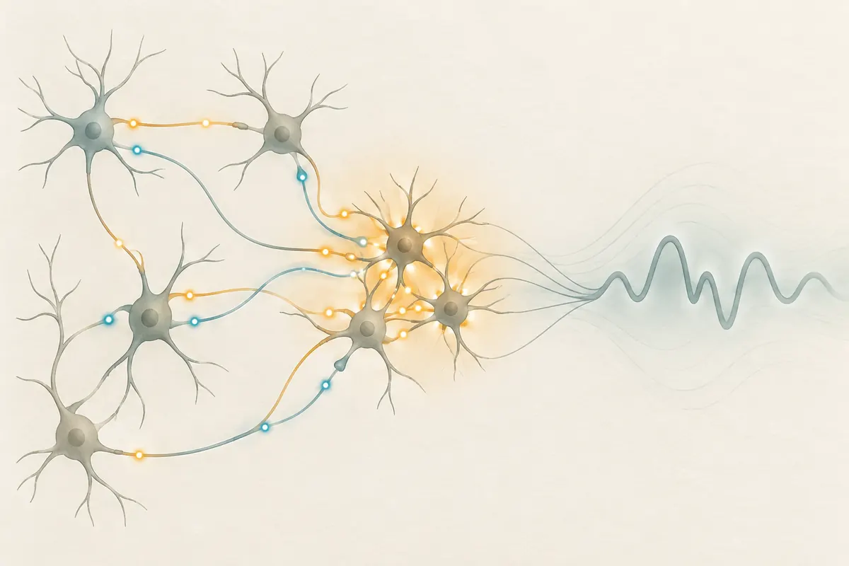
An interictal discharge occurs between clinical seizures; an ictal discharge accompanies seizure activity. A scalp EEG is not a direct microphone on one neuron. Each electrode records a combined electrical field from a broad region of tissue, shaped by the sources, their timing, and how the signal passes through the head. A waveform can therefore inform a network-level explanation without, by itself, establishing a person’s diagnosis, the cause of a symptom, or an individual prognosis.
Membrane excitability and synapses set the conditions for collective firing
Neurons integrate inputs through ion channels and synapses that alter membrane voltage. Glutamatergic excitatory input can move a cell toward firing, while GABAergic input often limits firing or shapes its timing. Yet the effect of inhibition depends on factors such as cell type, chloride gradients, developmental state, and circuit context. GABA signaling is not a simple universal brake, and glutamate is not a single switch that turns seizures on. The same transmitter can participate in different network dynamics under different conditions.
Synchronized activity is also part of ordinary brain function, including rhythms associated with sleep and information processing. Synchrony alone is not proof of disease. Concern arises when a network produces an abnormal pattern that becomes sufficiently large, sustained, or spatially organized to appear as epileptiform activity. Intrinsic bursting, recurrent excitatory connections, inhibitory interneuron timing, and local extracellular ion concentrations can all contribute. Their relative roles vary by brain region, seizure type, experimental preparation, and disease stage.
Interictal spikes and ictal discharges answer different questions
Interictal spikes are brief epileptiform events recorded between seizures. Ictal discharges are longer evolving patterns associated with a seizure. They may share some cellular or circuit features, but one should not assume they are interchangeable or that every interictal spike triggers a seizure. In some settings, synchronized inhibition can constrain activity; in others, excitatory pyramidal-cell bursts may contribute to the onset of an interictal-like event. The apparent role of a cell type depends on the signal being studied and the network’s state.
Hofer and colleagues studied human neocortical tissue obtained during surgery and maintained as slices. They distinguished lower-amplitude synchronous population activity, also seen in tissue without epilepsy, from more complex interictal-like epileptic discharges observed in epileptic tissue. Their recordings linked the asymmetric onset of those discharges to increased firing by excitatory cells, with bursting pyramidal cells a possible contributor.
The distinction between epileptogenesis and ictogenesis is equally important. Epileptogenesis is the longer process through which a brain may become more prone to spontaneous seizures after a predisposing factor or injury. Ictogenesis concerns how a particular seizure begins and develops. The processes are related, but evidence about one does not automatically settle the other. EEG patterns may help researchers investigate both, while their interpretation remains tied to the study design and clinical context.
EEG patterns and cellular recordings describe different levels
An interictal spike on an EEG is a pattern of electrical activity involving networks; it is not a direct recording of one neuron. Hofer and colleagues measured cell firing alongside local electrical signals in human tissue, but their slice experiment does not make every scalp waveform equivalent to a cellular event or establish what a pattern means for an individual. The method and experimental setting define what a result can support.
Specialist perspective: interictal activity has multiple mechanisms
In his specialist review of limbic networks and temporal lobe epilepsy, M. Avoli, affiliated with the Montreal Neurological Institute and McGill University describes several forms of interictal spikes and high-frequency oscillations. He notes that interictal events can arise through different mechanisms and may have different relationships to seizure generation. The review draws on human recordings as well as animal and in-vitro models, so its findings should remain tied to their studied context rather than treated as a universal account of epilepsy.