Atrial Fibrillation, Left Atrial Appendage Clots, and Stroke Risk
Atrial fibrillation can disrupt coordinated atrial contraction, allowing blood to pool and raising the possibility of clot-related stroke. This guide explains the left atrial appendage and why clinical risk assessment is not a score-only rule.
Publication & corrections: infostoa · Editorial standards
Individual author and specialist reviewer information is not provided for this article.
How an irregular rhythm can create a clot-related risk
Atrial fibrillation (AF) is an irregular heart rhythm in which the atria do not contract in a coordinated way. NHLBI explains that the upper and lower chambers may not work together normally, so the lower chambers may not fill completely or pump enough blood to the body. Blood may pool in the heart, increasing the chance of clot formation and stroke or other complications. A clot traveling through the circulation and blocking an artery supplying the brain is a possible route, not an inevitable outcome for everyone with AF. NHLBI also notes that AF may occur without symptoms, which can make it difficult to diagnose; symptoms alone cannot establish whether AF or a clot is present.
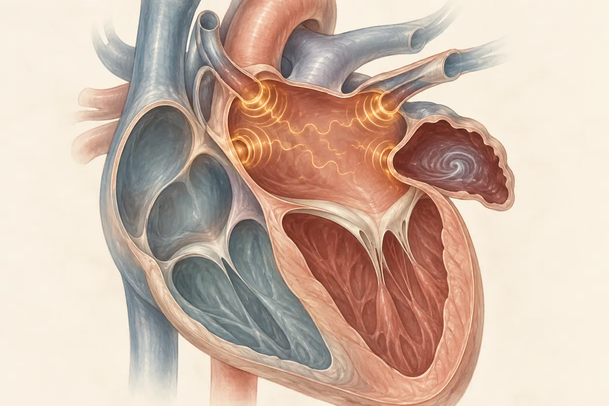
Why the left atrial appendage matters
The left atrial appendage (LAA) is a small pouch connected to the left atrium. In AF, altered atrial contraction and blood flow can make clot formation in the heart a concern; the 2023 ACC/AHA/ACCP/HRS guideline identifies the LAA as the most common site of thrombus formation in people with AF. This does not mean every clot forms there, that every person with AF has an LAA clot, or that the anatomy alone reveals an individual’s risk. Clinicians interpret imaging, symptoms, medical history, and other findings together. The useful takeaway is narrower: the LAA is a clinically relevant site to consider when specialists assess clot risk, while the presence or absence of a clot cannot be inferred from this general explanation.
A risk score is an input, not a treatment command
Clinical guidelines use validated tools to organize estimates of thromboembolic risk, but a score is not a stand-alone prescription. The 2023 guideline notes that estimated annual stroke risk can vary substantially among people with the same score and recommends evaluating stroke risk alongside bleeding risk and the rest of the clinical picture. Age, prior conditions, coexisting illness, bleeding factors, and a person’s circumstances all matter. That is why a threshold printed in a general article cannot tell a reader whether to start, stop, or choose a particular medicine or procedure. The score helps structure a clinician’s discussion; it does not replace that discussion.
Connect prevention decisions to a clinical record
NHLBI’s treatment guidance explains that anticoagulants reduce clots and stroke risk but also carry bleeding risk. Controlling the pulse and preventing clots are different goals: fewer palpitations are not a reason to stop prevention medicine on your own. A clinician assesses the need and choice.
Record the diagnostic test and date, symptom timing and duration, current medicine names, prior stroke or bleeding, other conditions and next review date. A fictional entry, “no palpitations today / previously confirmed AF / anticoagulation review planned,” does not mean “no clot risk.” Describe when and where bleeding occurred and any treatment. Neither a score nor bleeding history alone is a start/stop instruction.
Symptoms that should not wait for a log
As NHLBI’s stroke symptom guidance explains, sudden one-sided face or arm weakness or difficulty speaking warrants immediate local emergency help. Act even if symptoms disappear; do not wait for a risk calculation or routine appointment.